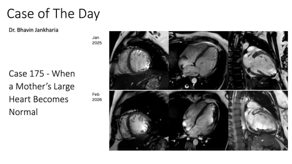
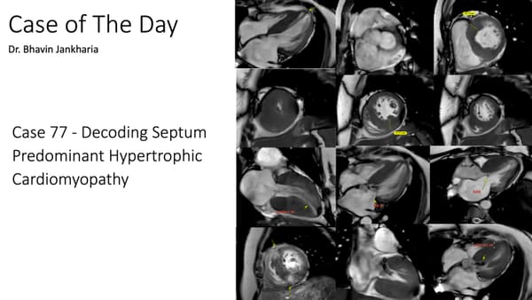
COD 175 - When a Mother’s Large Heart Becomes Normal
Peripartum cardiomyopathy is a complex condition where CMR has a role to play

Peripartum cardiomyopathy is a complex condition where CMR has a role to play

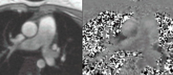
Cardiac MRI is an important tool to diagnose adult congenital heart disease including double-chambered RV
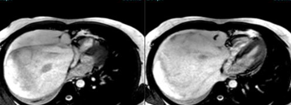
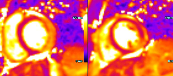
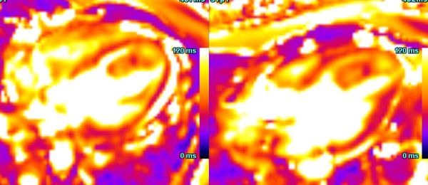
Cardiac MRI is a useful tool to diagnose amyloidosis. T1 mapping has become a mainstay, both native and ECV measurements. ECV measurement can now help quantify the amyloidosis burden and may help guide treatment based on how the ECV responds to specific treatment measures.
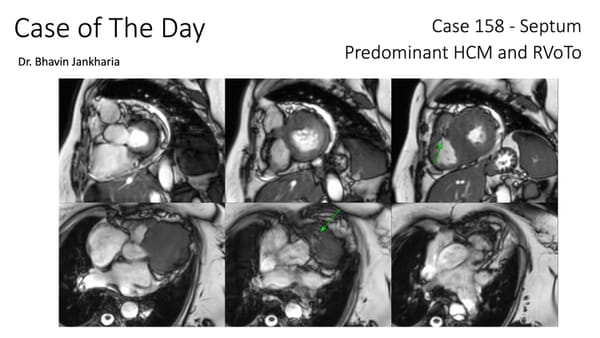
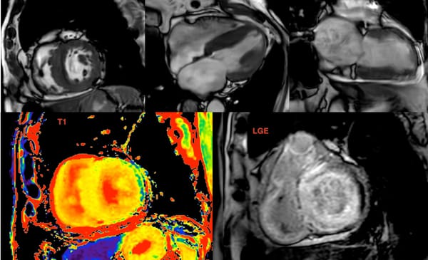
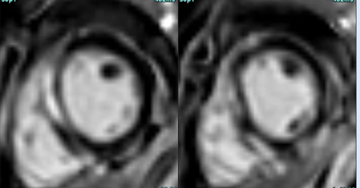
RVoTo involvement in HCM is uncommon but diagnosing it may change the management plan and prognosis
A common clinical presentation of LBBB induced cardiomyopathy is described in this post. It is always dilemma that if LBBB is cause or effect of dilated cardiomyopathy. However CMR can help in differentiating it from other causes and also in predicting response to device therapy. Utilise CMR to the fullest.
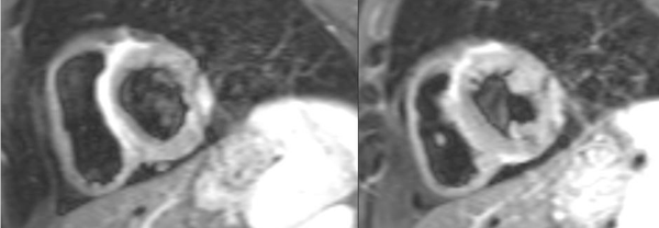
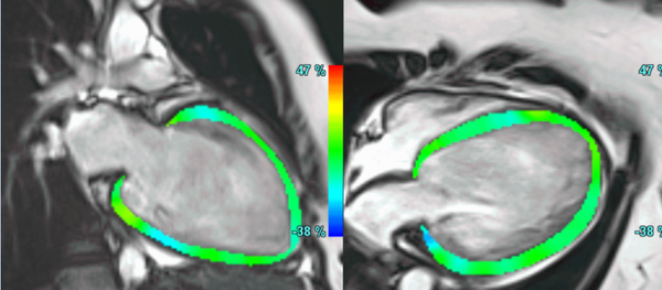
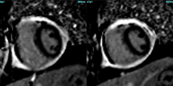
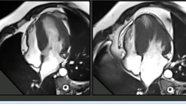
Understanding standard septum predominant HCM

Cardiac MRI does not show vegetations, easily especially if small. However, evaluating the transaxials through the valve may help pick up small nodules and to rule out mimics.

Infarcts
Diagnosing ventricular free wall rupture is not difficult, though these patients rarely come for cardiac MRI, since they are usually serious and clinically unstable.
Cardiomyopathy
Infarcts
Infarcts
Infarcts
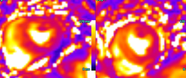
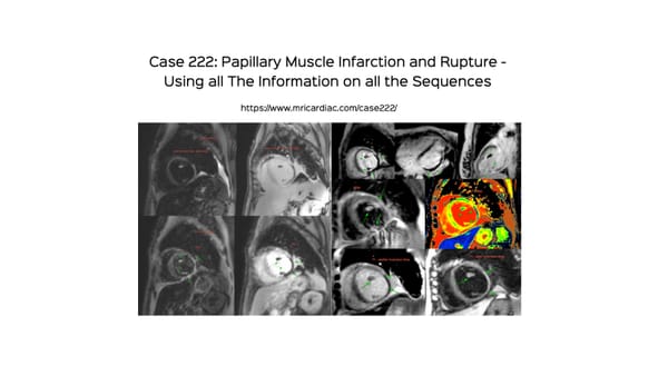
How to diagnose papillary muscle infarction and rupture using all the information we can get from all the routine sequences we obtain
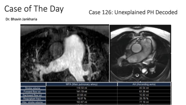
Pulmonary Hypertension
In patients with PH, radiologists can make a difference in identifying a few pathologies as causes of PH (leaving aside known cardiac and pulmonary diseases, where the conditions are already known). We should not miss these, especially shunts.

HCM
Cardiomyopathy
IMH
Hypertrophic Cardiomyopathy
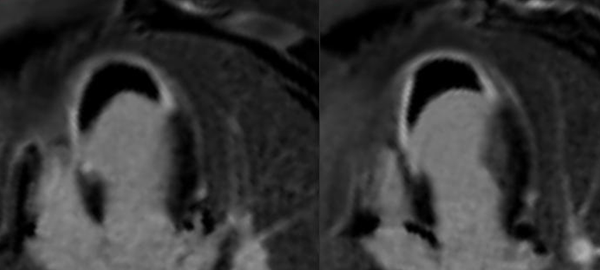
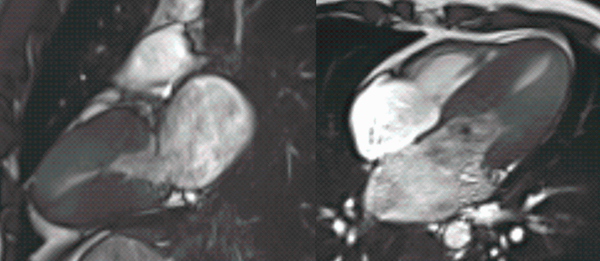
Ischemia in apical HCM appears to be universal and may be manifest at presentation or on follow-up.
Cardiomyopathy
Viability
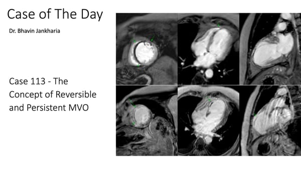
Persistent MVO carries a poor prognosis
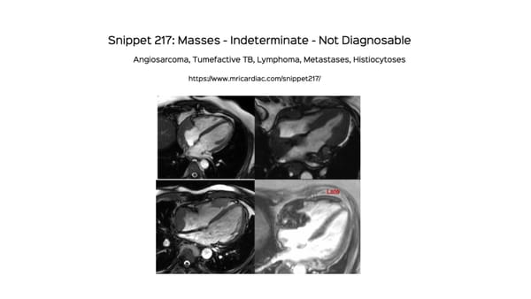
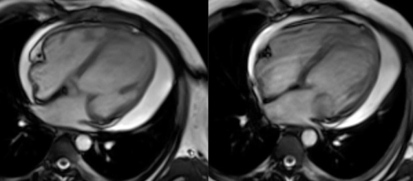
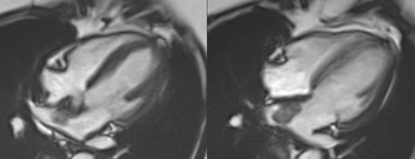
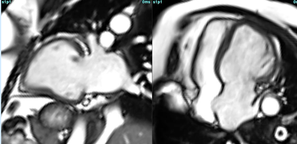
Tumors
A snippet on tumors that need a biopsy or either the tumor itself or of a related lesion for diagnosis.
Infections
Myxoma
Valvular HD