The DCRV Enigma
Cardiac MRI is an important tool to diagnose adult congenital heart disease including double-chambered RV
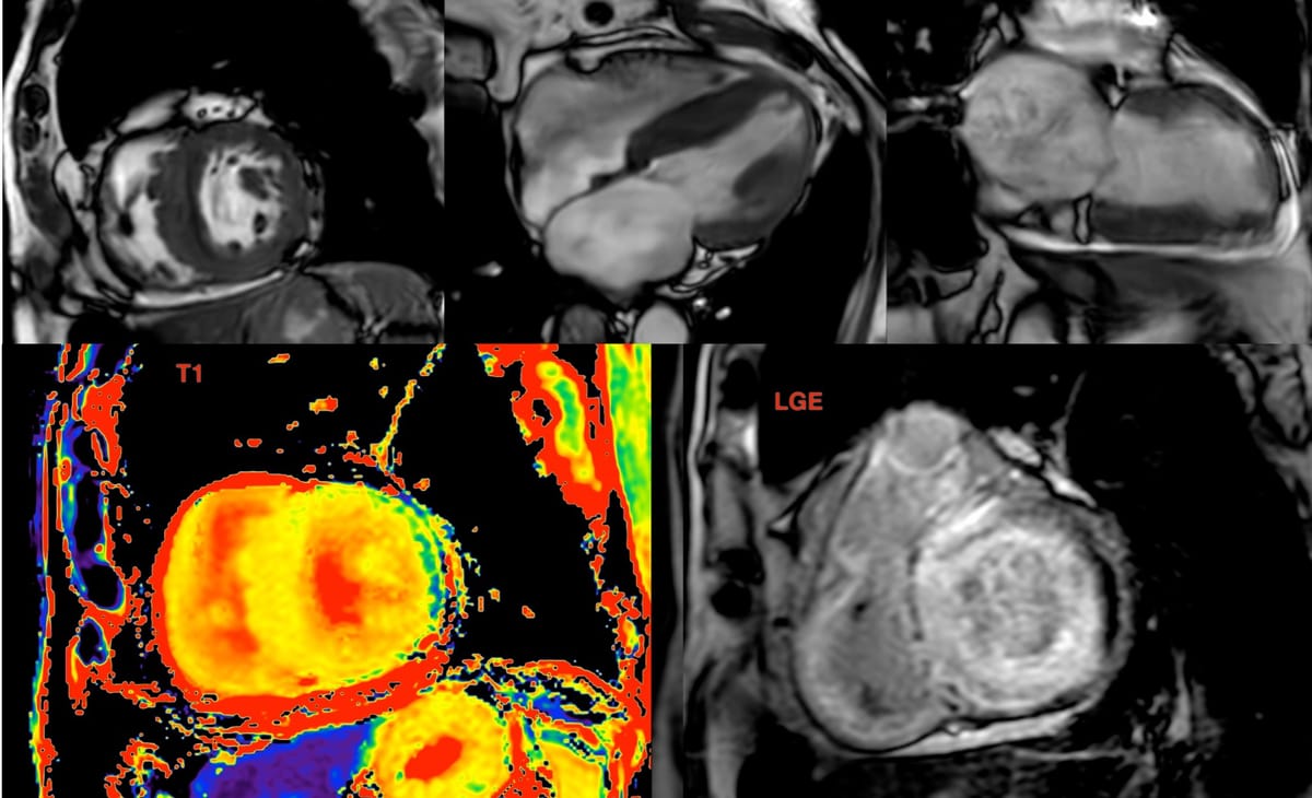
51-years old with giddiness every morning on waking up.
Echo showed RV changes.
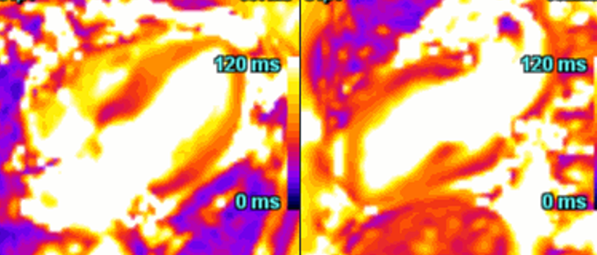
Cardiac MRI shows marked RV hypertrophy, the cause of which is abnormal musculature in the subpulmonic region, dividing the heart into a high-pressure proximal chamber and a low-pressure distal chamber, a double-chambered right ventricle or DCRV.
In this case, there are abnormal septo-parietal and septo-marginal trabeculae causing the stenosis.
To know each time a new post is up
Video
Related Case
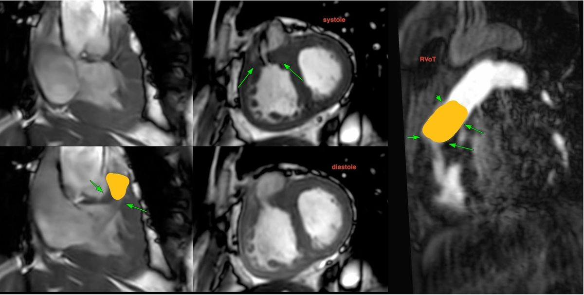
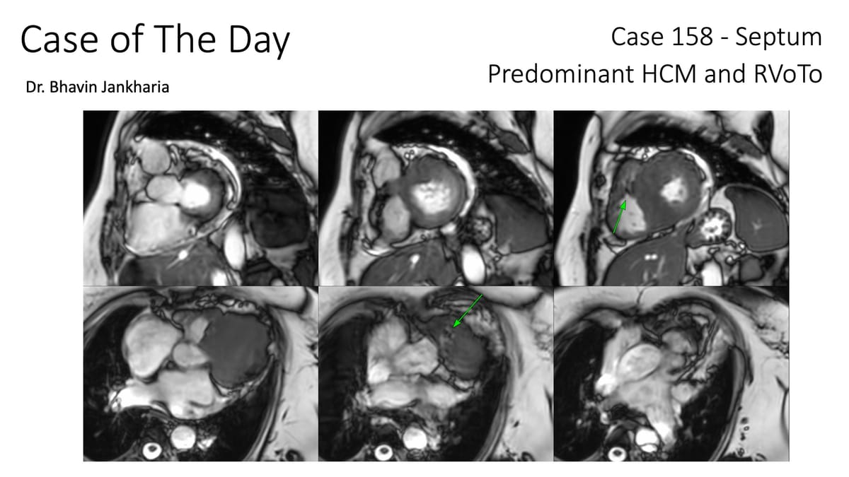
COD 158 - Septum Predominant HCM and RVoTo
RVoTo involvement in HCM is uncommon but diagnosing it may change the management plan and prognosis
Previous Post - Bhavin
The Value of Extracellular Volume (ECV) in ATTR - Transthyretin Amyloidosis
Cardiac MRI is a useful tool to diagnose amyloidosis. T1 mapping has become a mainstay, both native and ECV measurements. ECV measurement can now help quantify the amyloidosis burden and may help guide treatment based on how the ECV responds to specific treatment measures.
Previous Post - Priya
Map the heart!
Case based learning of Cardiac MRI