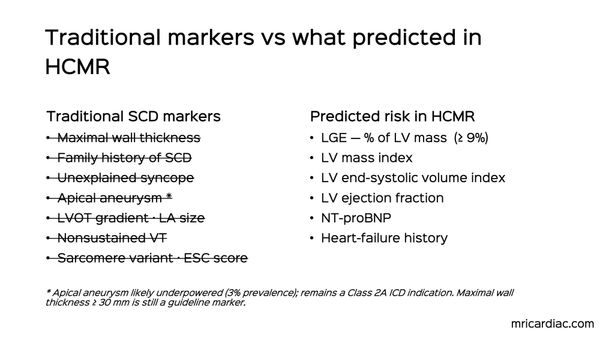
Multimodality Imaging of an LV Aneurysm
A complex long-standing, slow growing LV aneurysm
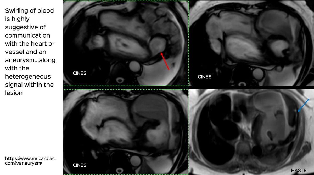
This 63-years old came with dyspnea. The X-rays of the chest, 12 years apart, showed a growing mass with eggshell calcification in the left hemithorax.
She was referred for a biopsy and eventually a diagnosis of an LV aneurysm was made. The video explains the way in which the diagnosis was made and the MRI findings.