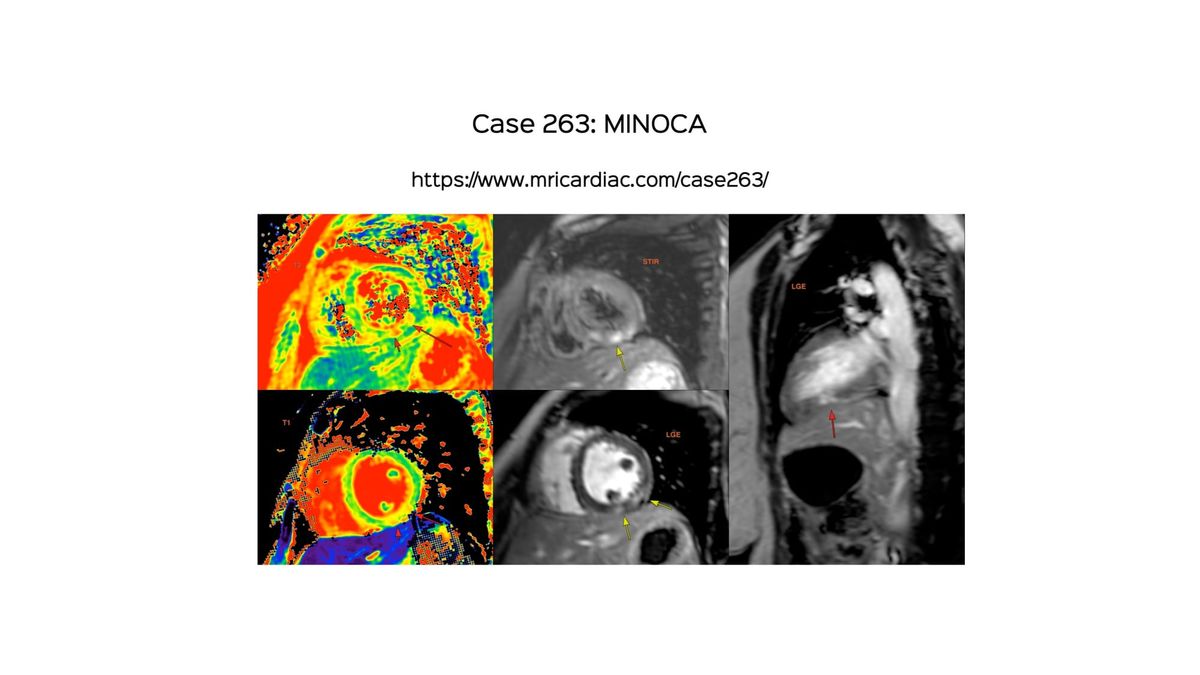
Case 263 - MINOCA
MINOCA needs to be understood well by radiologists and cardiologists, so that CMR, which is the cornerstone is performed at the earliest, rather than later.
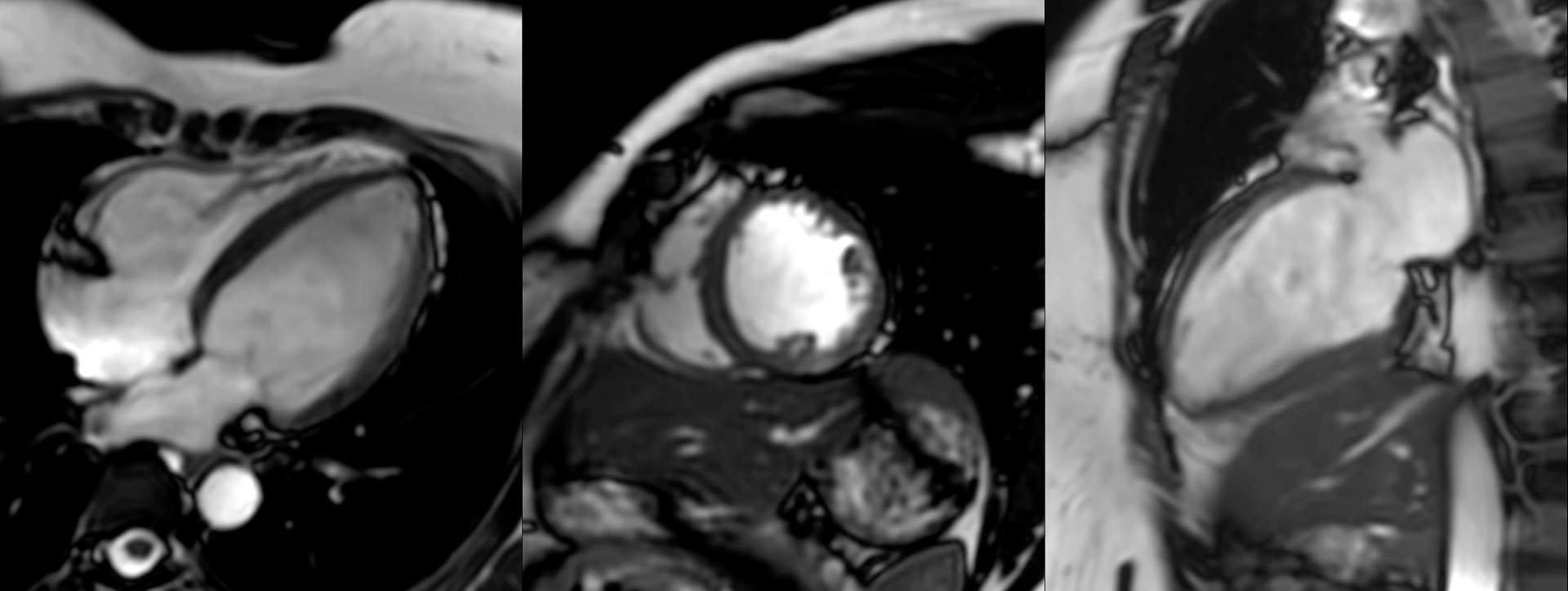
42-year-old had an acute cardiac event 7 days prior to the MRI with rising troponin over 48 hours, T-wave inversions and normal CT coronary angiogram.
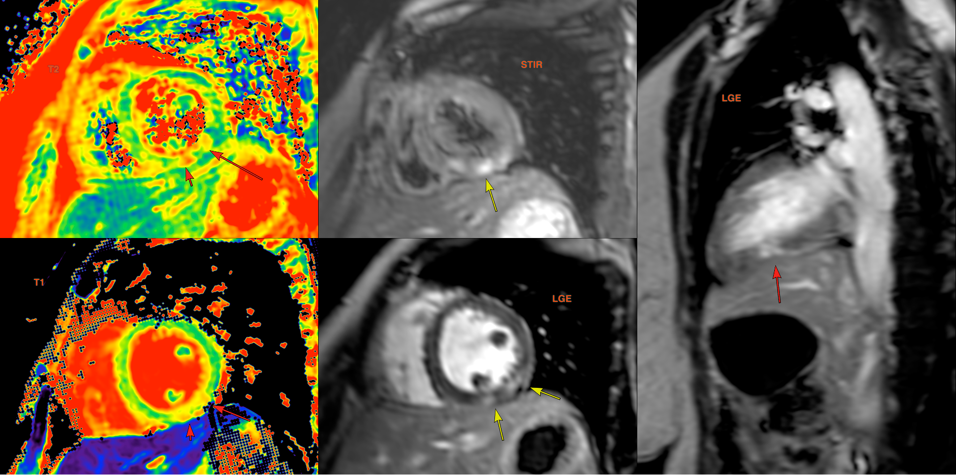
She was diagnosed to have MINOCA (Myocardial Infarction with Non-Obstructive Coronary Arteries) clinically.
The video describes the case, and specifically three recent papers that have changed the way we approach patients with MINOCA, ending with another case.
Previous Posts
Case 260 - Septal Flash
Septal flash on cine imaging can be further deconstructed with segmental strain imaging to help differentiate between LBBB cardiomyopathy and just a conduction abnormality.
 Bhavin Jankharia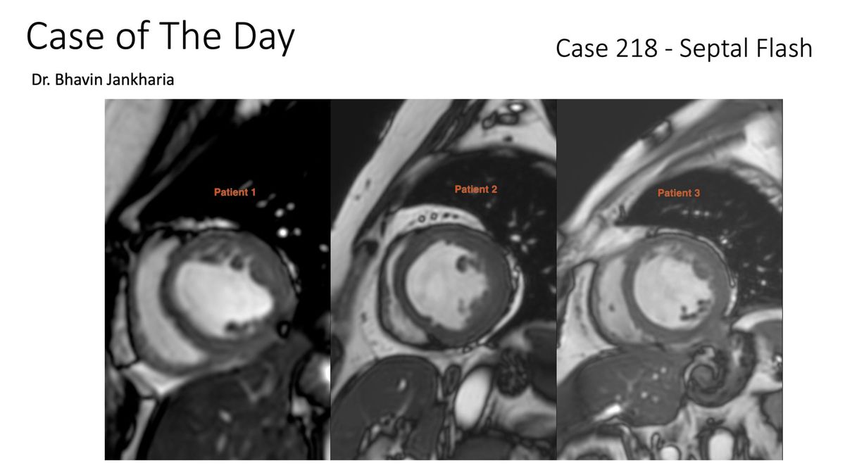
Previous Similar Posts
MINOCA Masquerade
Case based learning of Cardiac MRI
 Priya Chudgar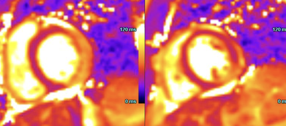
MINOCA Case
Case based learning of Cardiac MRI
 Priya Chudgar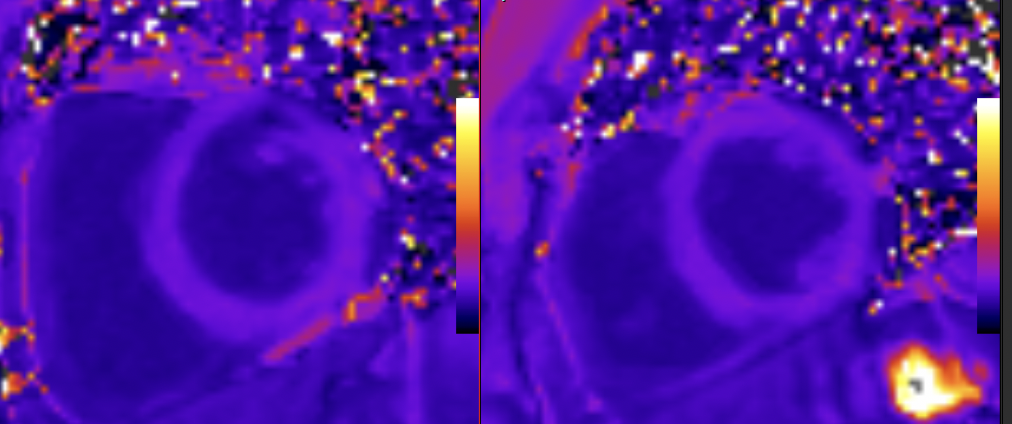
Case of the Day on YouTube
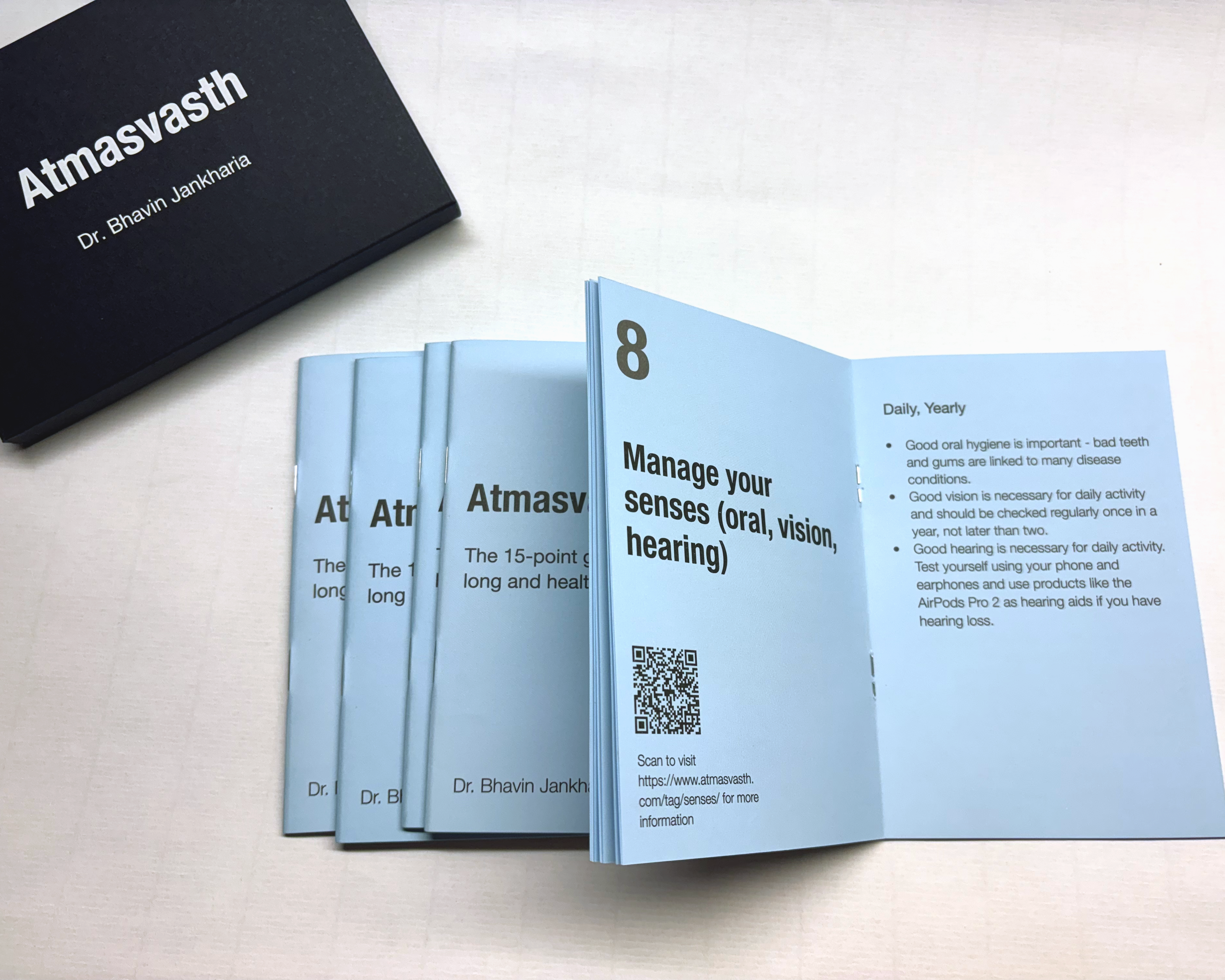
My New Atmasvasth Zine
The Atmasvasth 15-Point Guide to Living Long Healthy - Zine
This zine presents the 15-points in short, in a easy to digest and understand manner. 5 zines make a collection that comes with an envelope. The zines (also called booklets in the past) make great gifts. There are 5 purchase options. Selection Expected checkout 1 pack ₹300 + ₹50 shipping = ₹350 2 packs ₹550 + ₹50 ship
 JPLLP
JPLLP