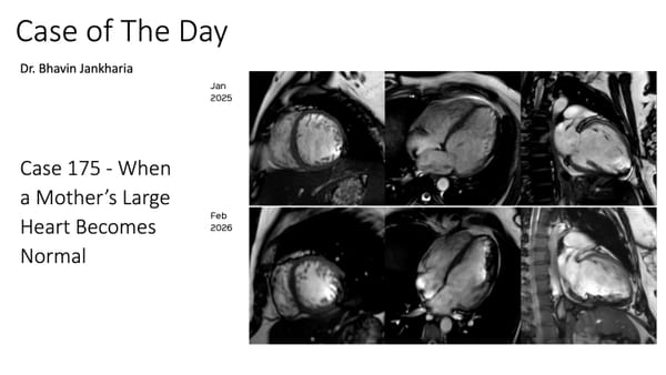
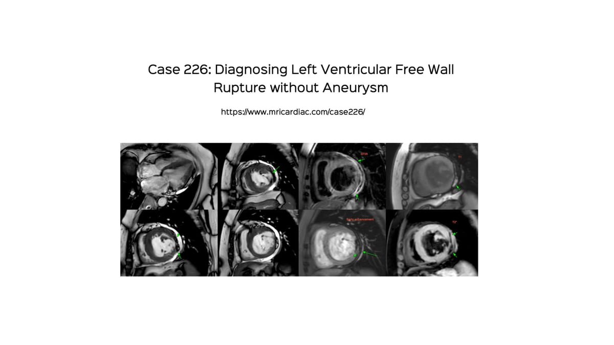
Case 226: Diagnosing Left Ventricular Free Wall Rupture without Aneurysm
Diagnosing ventricular free wall rupture is not difficult, though these patients rarely come for cardiac MRI, since they are usually serious and clinically unstable.
69-years old with acute coronary syndrome (ACS), 4-5 weeks prior to the cardiac MRI with a coronary angiogram 3 weeks prior to the cardiac MRI showing triple vessel disease.
She was stable and stent for a viability study.
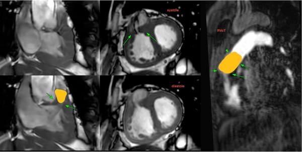
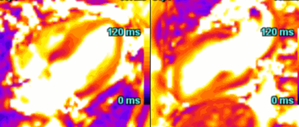
CMR showed a lateral wall infarct with hemorrhage and other features best appreciated in the video which describes the case and the various types of free wall ruptures with additional cases.