Case 195: Looking for and Diagnosing Tuberculosis in the Lungs When CMR Findings are Classic for Sarcoid Cardiomyopathy
Every patient with suspected sarcoid cardiomyopathy must be thoroughly investigated to make sure it is not TB masquerading as sarcoid
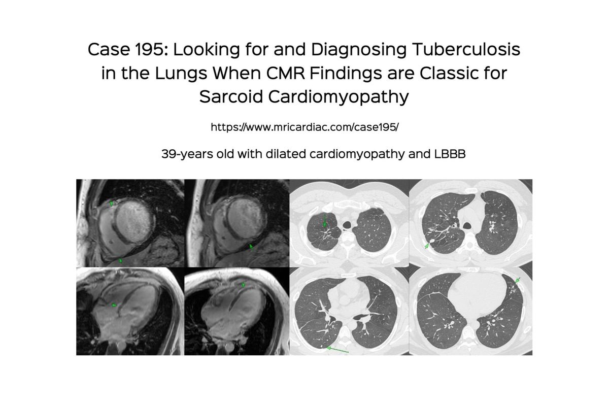
39-years old presented to us with a diagnosis of dilated cardiomyopathy on echo on left bundle-branch block (LBBB) on ECG.
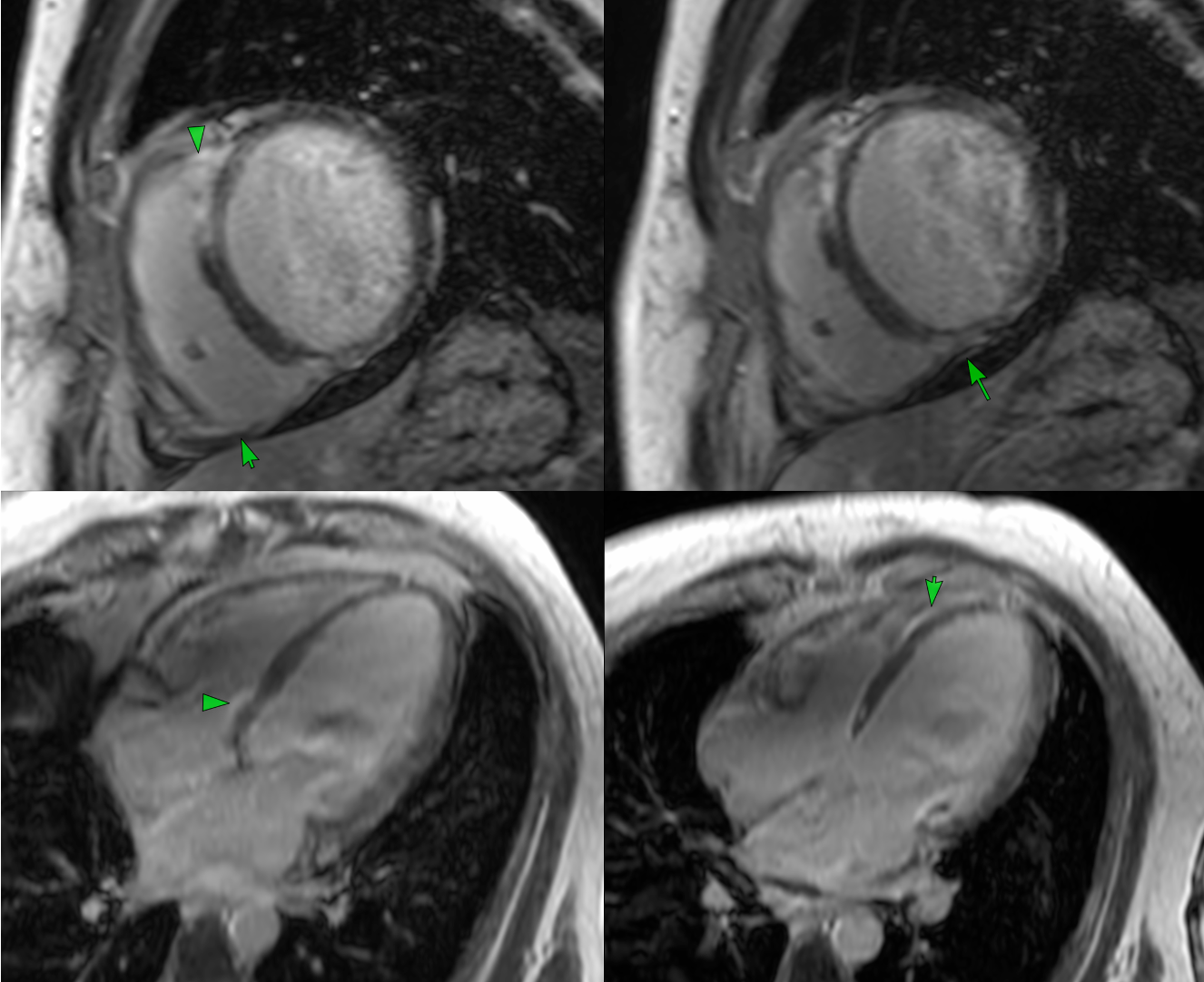
There was biventricular systolic dysfunction with inferior wall thinning.
The late Gd images showed epicardial, septal, mutifocal and RV enhancement, typical of sarcoid cardiomyopathy.
The diagnosis was finally obtained on the lung images of a whole body PET/CT and then bronchoscopy and lavage.
The video below explains the case and the importance of looking at the lungs and the rest of the body to make the correct diagnosis in a suspected granulomatous cardiomyopathy.