

Latest

Revisiting restriction

Follow the flow..!
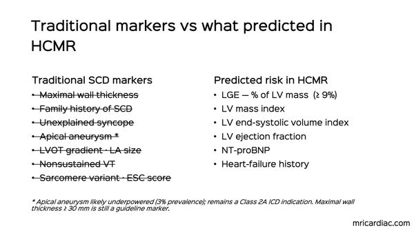
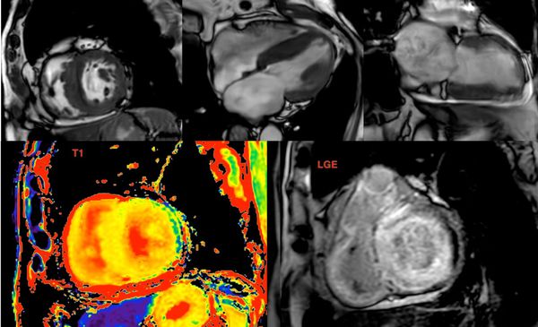
Case 255 - The New HCM Risk Predictors - What Matters and What Doesn't
CMR is central to HCM management. Merely reprinting values does not help. Flag the ones that matter, LGE%, LV mass index, LV end-systolic volume index, end-diastolic wall thickness > 30 mm and apical aneurysm.

Different faces of HCM

Look beyond the obvious!

Understanding SAM

Arrythmogenic cardiomyopathy
Case 250 - The Banana-Shaped Heart
MRI and CT are both useful and complimentary when it comes to pericardial disease

Mitral annulus disjunction

Cardio-embolic stroke

Peripartum Cardiomyopathy
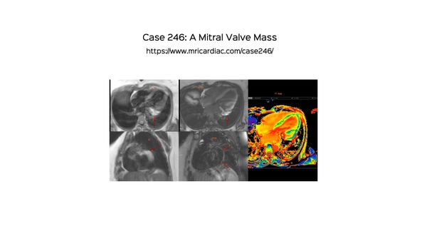
Case 246 - A Mitral Valve Mass
A tumor protocol cardiac MRI often allows a definite diagnosis to be made as in this case.

Pericardial cyst

True LV aneurysm

ARVC
Adult Congenital
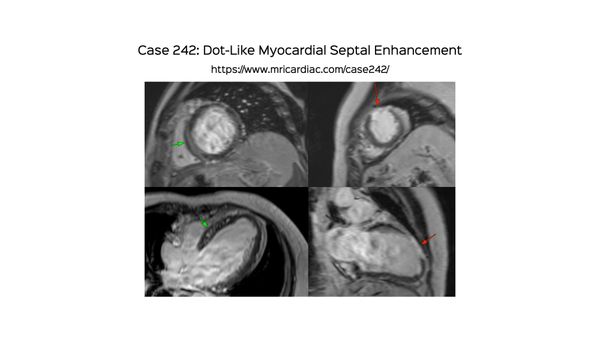
Case 242 - Dot-Like Myocardial Septal Enhancement
An unusual pattern of dot-like enhancement on 4C in the septum should alert us to look for this condition on CMR (prominent vessels, collateral vessels, infarcts) and altered patterns of perfusion and ischemia on stress CMR

CMR Shorts
Many faces of myocarditis

LGE
Fat in the scar

Amyloidosis
Cardiac Amyloid
Peripartum
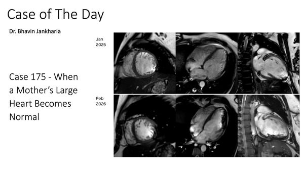
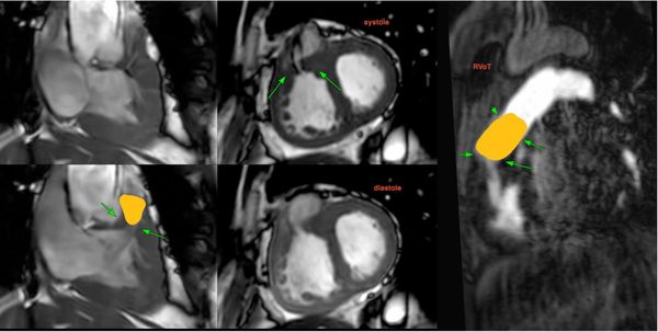
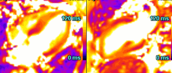
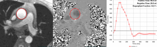
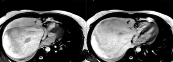
COD 175 - When a Mother’s Large Heart Becomes Normal
Peripartum cardiomyopathy is a complex condition where CMR has a role to play

Non-Ischemic Cardiomyopathy
Case series: Genetic CMP
Adult Congenital
The DCRV Enigma
Cardiac MRI is an important tool to diagnose adult congenital heart disease including double-chambered RV
Cardiomyopathy
Map the heart!
Mitral Valve
Double valve
Amyloidosis
The Value of Extracellular Volume (ECV) in ATTR - Transthyretin Amyloidosis
Cardiac MRI is a useful tool to diagnose amyloidosis. T1 mapping has become a mainstay, both native and ECV measurements. ECV measurement can now help quantify the amyloidosis burden and may help guide treatment based on how the ECV responds to specific treatment measures.
Tumors
The Heart Under Siege !
Cardiomyopathy
MINOCA Masquerade
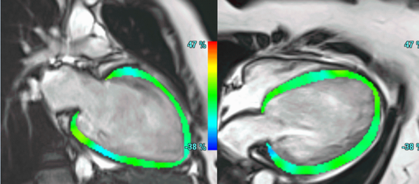
Hypertrophic Cardiomyopathy
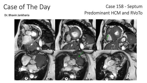
COD 158 - Septum Predominant HCM and RVoTo
RVoTo involvement in HCM is uncommon but diagnosing it may change the management plan and prognosis
Cardiomyopathy
Not in Sync anymore..!
A common clinical presentation of LBBB induced cardiomyopathy is described in this post. It is always dilemma that if LBBB is cause or effect of dilated cardiomyopathy. However CMR can help in differentiating it from other causes and also in predicting response to device therapy. Utilise CMR to the fullest.
Cardiomyopathy
Hope it fades..!
Hypertrophic Cardiomyopathy
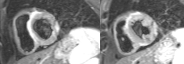
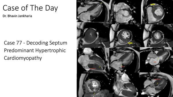
COD 077 - Decoding Septum Predominant Hypertrophic Cardiomyopathy
Understanding standard septum predominant HCM