The Value of Extracellular Volume (ECV) in ATTR - Transthyretin Amyloidosis
Cardiac MRI is a useful tool to diagnose amyloidosis. T1 mapping has become a mainstay, both native and ECV measurements. ECV measurement can now help quantify the amyloidosis burden and may help guide treatment based on how the ECV responds to specific treatment measures.
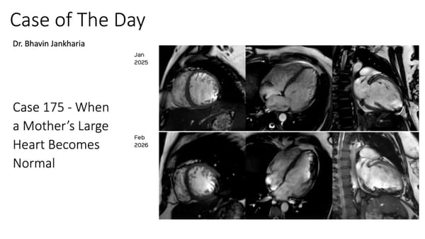
83-years old presented with HFrEF in June 2025, echo showing LVH with reduced EF.
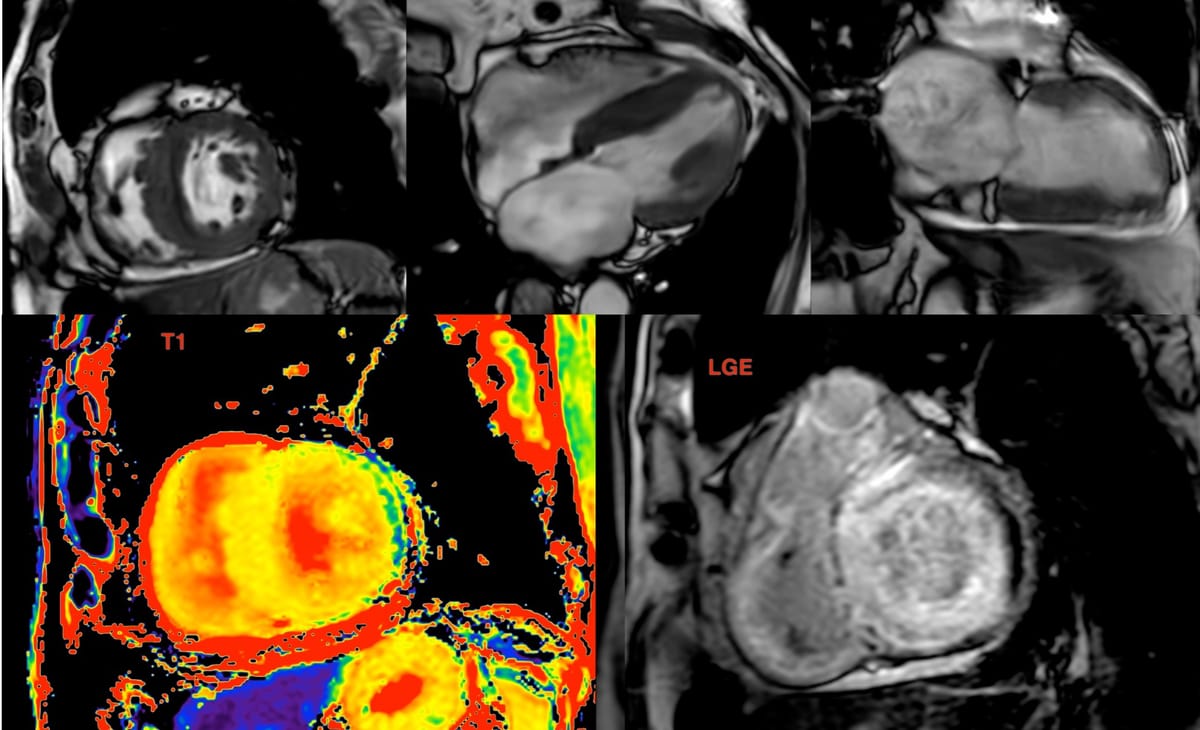
Cardiac MRI shows typical findings of amyloidosis with diffuse transmural enhancement, deranged contrast kinetics and raised T1 and an ECV of 60%.
The video discusses the case and the importance of T1 and ECV, specifically based on a new JACC article that I read yesterday (Sheikh A et al - JACC 2026 Vol 87, pg 505), published a day prio that discusses the importance of ECV in quantifying transthyretin amyloidosis.
To know each time a new post is up